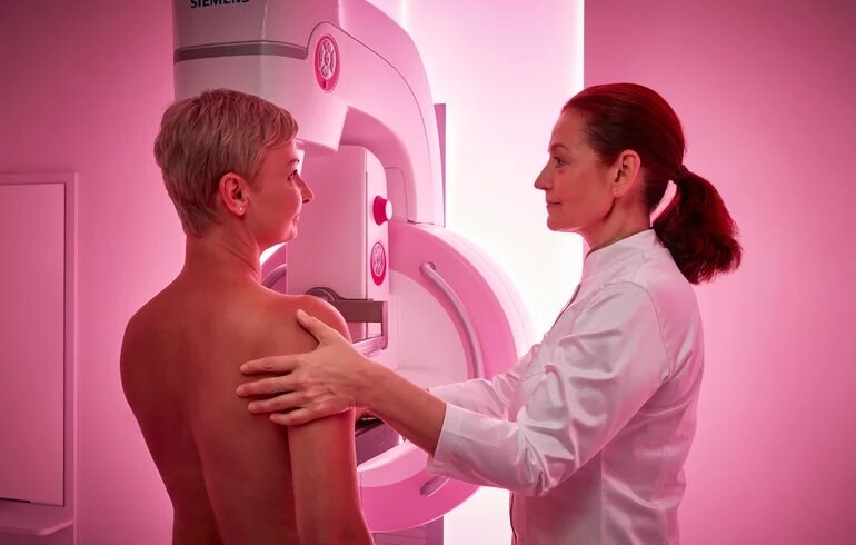
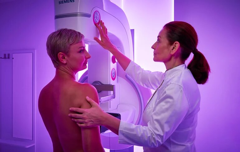
Mammography is an x-ray examination of the breasts that uses a low dose of radiation to detect changes in breast tissue, such as tumors or calcifications.
Mammography
The examination is recommended during the first half of the cycle, i.e., within 15 days after the first day of menstruation. During the examination, the breasts are compressed from two directions and images are taken.
The mammography examination is performed using SIEMENS Healthineers' top-of-the-range Mammomat Inspiration digital 3D mammography machine, which provides uncompromising image quality with up to 30% reduction in radiation dose. The device's special, relaxing light effects help patients relax, ensuring thorough examination and optimal image results.
At Dr. Rose Private Hospital, breast cancer screening is performed as part of a complex breast diagnostic examination. As part of the process, a two-way mammography x-ray is taken of both breasts, after which the radiologist – who specializes in breast diagnostics – immediately examines the patient. The examination also includes taking a detailed family and individual medical history, a physical breast examination, and a breast ultrasound. After summarizing the results, the radiologist may also recommend tomosynthesis if necessary. Tomosynthesis is a modern breast diagnostic method that uses x-rays, as in traditional mammography, but takes images from multiple directions. From these, the system creates a layer-by-layer, three-dimensional image of the breast, so that pathological changes – especially in the case of dense breast tissue – can be detected with even greater accuracy. Since the examination involves a higher radiation dose than standard mammography, it is recommended only in justified cases.
If necessary, biopsy (fine and thick needle) and stereotactic sampling are also available.


