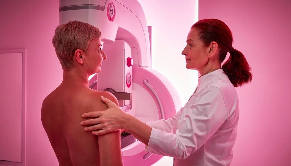
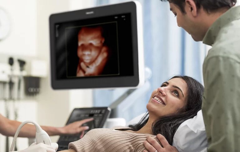
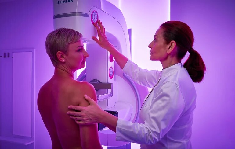
We use a state-of-the-art direct digital mammography machine. If required, a three-dimensional tomosynthesis, biopsy (thin and thick needle) and stereotactic sampling are also performed. Mammography is performed with the SIEMENS Healthineers’ high-end Mammomat Inspiration, which delivers uncompromised image quality with up to 30% less radiation dose. Ultrasound examinations are performed with the Philips Affiniti 50, which provides reliable performance, high-quality images and rapid diagnostic information. At Dr. Rose Private Hospital, mammography examinations follow national and international protocols.
The use of imaging modalities in the advanced screening of breast tumors
Mammography
Mammography is recommended in cases of symptoms or complaints over the age of 30-35 years – except if such an examination has been done within the last year and there is no reason to repeat it, e.g., a new symptom of a suspected tumor. It can be performed on women under the age of 30 if justified, with the addition of targeted, magnified images and guided sampling if necessary. Mammography is the only scientifically proven method for screening women at average risk to reduce breast cancer mortality.
Tomosynthesis
Digital 3D Breast Tomosynthesis (DBT) is a screening procedure based on 2D full-field digital mammography (FFDM), in which an x-ray tube moves in an arc around the breast to produce 10-15 overlapping digital images of the breast at low radiation doses in a short time. The data set is processed by computer to produce multiple 2D thin-slice images and reconstructed 3D images similar to conventional synthetic 2D overview images. To reduce the radiation dose, it is recommended to replace part or all the conventional 2D images with the synthetic 2D images, if the device has an official certificate (e.g., FDA approval). 3D tomosynthesis is more sensitive in the assessment of breast structure, and hidden lesions are easier to detect due to this higher sensitivity. Tomosynthesis is effective in clarifying the overlapping tissues (summation shadow) that pose diagnostic difficulties in conventional 2D imaging. By analyzing thin-slice images, breast structure can be evaluated without the confounding effect of summation shadows, allowing a more accurate assessment of abnormal structural distortions and lesion boundaries, and eliminating false-positive results due to summation shadows. This results in a 29-41% increase in tumor detection, a significant reduction in the recall rate when used in screening, and avoidance of unnecessary biopsies. In breast screening, tomosynthesis is particularly advantageous for breast structures (dense fibrotic, fibroadenotic) where conventional mammography has a lower sensitivity.
Mammography-guided stereotactic sampling
Mammography-guided stereotactic sampling is necessary for non-palpable lesions that cannot be identified via ultrasound and are not definitely benign, e.g., microcalcifications. Targeting can be done in sitting/supine/lying on one side positions. Only lesions visible via tomosynthesis, most often structural distortions, can be targeted by tomosynthesis-guided stereotactic biopsy.
Ultrasound-guided sampling
Ultrasound-guided sampling of the breast and regional lymph nodes is recommended when palpable or non-palpable lesions are clearly visible via ultrasound examination.
Reference: our article is based on the technical guidelines of the IV. Hungarian Breast Cancer Consensus Conference.


