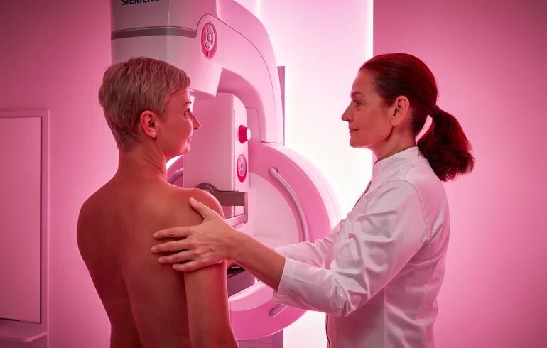
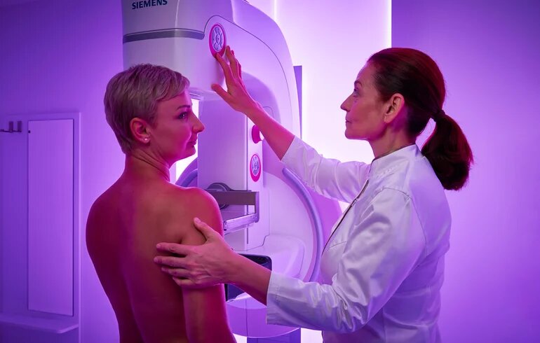
At Dr. Rose Private Hospital, breast cancer screening is performed with a complex breast diagnostic examination. During the examination, a two-dimensional mammography x-ray of both breasts is taken, and then the patient is immediately examined by a radiologist experienced in breast diagnostics. The procedure includes taking a detailed family and individual medical history, a physical breast examination, and a breast ultrasound. After comparing the results, the radiologist will also recommend a tomosynthesis examination if necessary. This examination protocol ensures the highest accuracy and the fastest results, without unnecessary radiation exposure.
Complex breast screening
Elements of the examination
- Mammography: an x-ray examination of the breasts that uses a low dose of radiation to detect changes in breast tissue, such as tumors or calcifications.
- Family and individual medical history: the doctor will ask the patient in detail about their own and their family's health history, for example, whether close relatives have had breast cancer or other cancers, whether they have had breast disease, surgery or hormone treatment in the past, and any lifestyle factors (e.g., number of births, time of menopause) that may influence the risk.
- Physical examination: During a breast examination, the doctor will manually examine the breasts and armpits to feel for any changes, such as lumps, hardness, or tender areas.
- Breast ultrasound: a painless, radiation-free test that uses soundwaves to provide an image of breast tissue. It can be used to detect cysts, lumps, and other abnormalities.
- If necessary, tomosynthesis: tomosynthesis is a modern breast diagnostic procedure that uses x-rays, as in traditional mammography, but takes multiple images from multiple directions. From these, the machine creates a layer-by-layer, three-dimensional image of the breast, which allows for even more accurate detection of pathological changes, especially in dense breast tissue. The examination involves higher radiation exposure than mammography, hence it is only recommended in justified cases.
The results are immediately evaluated by the radiologist. Depending on the results and the patient's individual condition and risk factors, further tests may be recommended, if necessary, such as a biopsy or stereotactic sampling.

