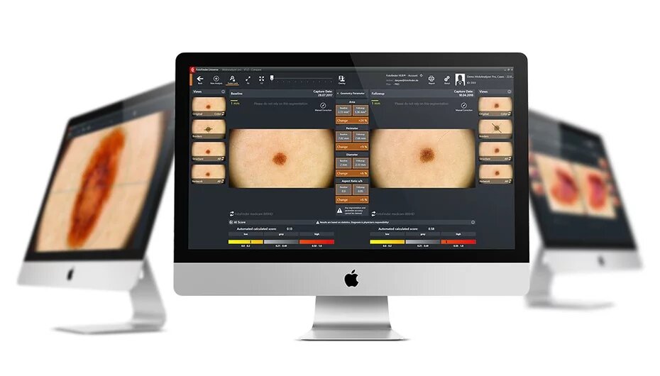
With this innovative technology, even the smallest changes in moles can be accurately tracked. By comparing moles recorded in full high-definition resolution at different times, any possible malignancy can be determined even more accurately. During the examination, in addition to the moles, the affected areas of the body are also recorded, thus allowing even more thorough long-term observation of the moles and any new skin formations.
During the examination, we first take overview images of the skin. The videodermatoscope is then used to take microscopic images of any atypical moles, which can be evaluated and analyzed. Digital storage of skin images allows for an objective comparison of past and current examinations during regular inspections, so even the smallest changes are detected.
Preventing skin cancer with FotoFinder®
- long-term and complete documentation of the entire skin surface and all moles
- examination of the whole body and initial analysis of skin lesions
- regular examinations detect new and changed moles at an early stage
- less unnecessary mole removal
The high-risk group comprises
- fair skin type, sensitive to sunlight
- a large number of moles
- atypical or altered moles
- if you have had severe sunburn in your childhood or in young adulthood
- if you have a history of skin cancer in your family
- if you have had a skin tumor before
- skin damage and/or exposure to increased sunlight because of an occupation
What should we pay attention to?
Be sure to tell your doctor if your moles show the following changes:
- you notice a sudden or gradual change
- irregular shape
- blurred, irregular edge
- non-uniform color
- size is greater than 5mm, or has increased
- protrusion or uneven surface
- freshly formed mole
- bleeding mole
- the environment of the mole has changed, e.g., redness, whitening, swelling
- discomfort: itching, burning, foreign body sensation
It is important for the effectiveness of dermatological screenings that everyone be examined once a year so that we can recognize malignant changes in time and provide a surgical solution as soon as possible.
